How Does the Shape of Orchids Relate to Gene Expression?
The shape of an orchid flower reflects which developmental genes are active, where they are active, and which proteins they form complexes with. Different combinations of gene expression tell floral tissues to become sepals, petals, the specialized lip, or the reproductive column.
This is not a case where one gene directly draws one flower shape. Orchid form emerges from a regulatory program involving duplicated genes, tissue-specific expression, protein interactions, and growth over time.
How does the shape of orchids relate to gene expression?
Orchid shape is produced by spatial gene expression. Cells in each floral region read different combinations of regulatory genes, then make proteins that direct organ identity, symmetry, pigmentation, and growth.
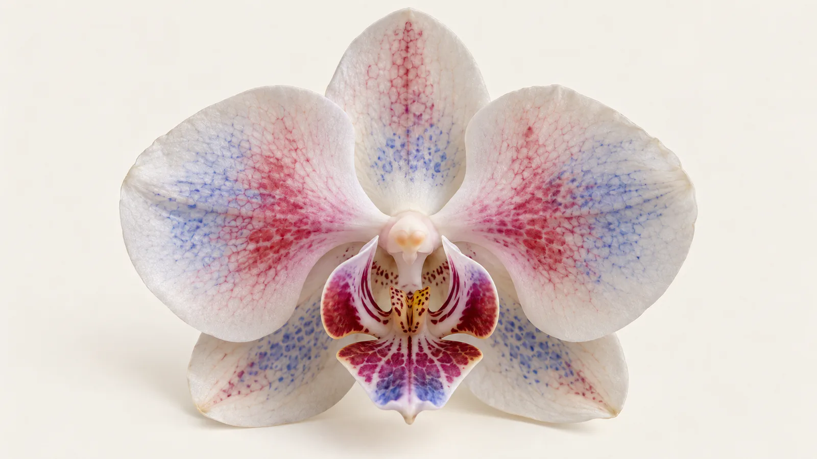
An orchid flower usually has three outer sepals, two inner petals, one highly modified petal called the lip or labellum, and a central column that combines male and female reproductive structures.
These organs begin as small groups of similar cells. Their final differences appear because the cells do not express the same developmental genes at the same levels or times.
Many of the key regulators are MADS-box transcription factors. A transcription factor binds DNA or works in a regulatory complex to influence which other genes are transcribed.
The resulting downstream program changes cell division, expansion, polarity, pigment production, and tissue structure. Those cellular changes become the visible architecture of the flower.
What genes control orchid flower shape?
The best-studied orchid shape regulators belong to the MADS-box family, especially AP3/DEF-like, PI/GLO-like, and AGL6-like genes.
These genes are related to the class B and related floral identity genes described by the ABC model of flower development. Orchids expanded and modified this toolkit through gene duplication.
Duplicated genes can keep some ancestral functions while acquiring new expression patterns or protein partners. That gives evolution more regulatory combinations without requiring an entirely new molecular system.
In Phalaenopsis, related AP3/DEF-like genes show different expression across outer tepals, inner petals, the lip, and the column. The pattern is combinatorial rather than one gene per organ.
A major review of orchid floral development describes how these expression patterns support an orchid-specific extension of the ABC model called the orchid code. Read the open-access review.
| Floral feature | Regulatory idea | Visible result |
|---|---|---|
| Petaloid sepals | Class B activity extends into the outer whorl | Sepals resemble colorful petals |
| Two lateral petals | A sepal or petal identity complex dominates | Bilaterally arranged inner tepals |
| Lip or labellum | A distinct combination of AP3/DEF-like and AGL6 proteins forms | One petal develops a specialized shape |
| Column | Reproductive organ programs are reorganized and fused | Stamens and pistil form one central structure |
| Bilateral symmetry | Gene activity differs across floral axes | One side or organ develops differently from another |
What is the orchid code?
The orchid code is a model in which combinations of related floral regulators specify sepals, petals, and the lip.
Its central idea is that organ identity depends on protein complexes. Different AP3/DEF-like proteins combine with PI/GLO-like and AGL6-like partners, creating regulatory complexes with different targets.
Researchers often summarize two important combinations as SP complexes for sepal and petal identity, and L complexes for lip identity.
Functional experiments reported in Nature Communications support this division. Changing B-class and AGL6 gene functions can shift tissue between lip-like and sepal or petal-like identities. See the study.
This explains why closely related organs can look radically different. They share much of the same genetic toolkit, but their cells assemble different regulatory combinations.
Gene expression is a recipe, not a blueprint
A blueprint suggests that every shape is drawn directly in DNA. A better model is a recipe: regulatory proteins alter other genes, cells respond to local signals, and tissue growth turns those instructions into form.
Why do orchid sepals look like petals?
Orchid sepals look petal-like because petal identity gene activity extends into the outer floral whorl.
In many flowering plants, sepals are green protective organs and class B gene activity is mainly associated with petals and stamens. In orchids, expanded B-class expression helps give all six tepals a petaloid appearance.
The result is a flower with three colorful outer sepals and three inner petals, although one inner petal becomes the lip.
This shared petaloid program does not make every organ identical. Additional expression differences split the lip from the other petals and shape each organ along its own developmental trajectory.
Why is the orchid lip different from the other petals?
The lip is different because it activates a distinct regulatory complex that redirects petal development.
Lip development changes more than outline. It can alter cell shape, ridges, hairs, pigments, scent, and nectar-related structures. These traits help guide or position pollinators.
Spatial transcriptomic work has detected distinct expression domains for MADS-box and other regulatory genes across orchid lip, column, and tepal tissues. See the spatial study.
The lip therefore illustrates a key rule of developmental biology: organs can share an evolutionary origin while diverging because their gene regulatory states differ.
What do peloric orchids reveal?
Peloric orchids reveal the gene-shape connection because their unusual symmetry often accompanies altered floral identity gene expression.
A typical orchid is bilaterally symmetrical. A peloric flower may develop three lip-like petals or replace the lip with a petal-like structure, making the flower more radially symmetrical.
Those transformations are informative because they change organ identity, not merely organ size. If reducing or relocating a lip-associated expression program converts one organ toward another, the program is likely involved in specifying that identity.
Researchers test this relationship with expression assays, mutant or transgenic experiments, protein interaction studies, and comparisons among normal and peloric flowers.
Expression alone shows correlation. Functional perturbation provides stronger evidence because it asks whether changing the regulator changes the organ.
Is orchid shape controlled by simple Mendelian inheritance?
Some visible orchid traits can segregate as major-gene traits, but overall flower shape is usually not controlled by one simple dominant or recessive allele.
Shape depends on multiple regulators, their expression control regions, protein interactions, hormone responses, and downstream growth genes. Environmental conditions can also affect organ size and development.
A mutation in one major regulator can produce a dramatic transformation. That does not mean the normal shape was made by that regulator alone.
The distinction is important. A single mutation may disrupt a network at a high-leverage point, while the network still contains many necessary components.
How do scientists measure gene expression in orchid flowers?
Scientists compare RNA abundance across organs, stages, cell types, and flower variants to identify genes associated with each shape.
RT-qPCR can test a small set of candidate genes. RNA sequencing measures thousands of transcripts at once, while spatial transcriptomics preserves information about where expression occurs inside the organ.
A basic experiment might collect outer sepals, lateral petals, lip, and column from matched flowers. Researchers then identify genes whose expression differs by organ and test enriched regulatory or developmental functions.
Bulk RNA-seq averages many cells. A lip sample can contain epidermal, vascular, secretory, and structural cells, so an average may hide small but important expression domains.
Spatial methods and in situ hybridization address that limitation by showing where transcripts occur. Protein interaction assays test whether the encoded regulators can form the complexes proposed by the orchid code.
The strongest studies combine these evidence types. Expression locates candidates, interaction data suggests mechanism, and perturbation tests causality.
Does higher expression always mean a larger orchid organ?
No. Higher expression does not automatically produce a larger organ or a stronger trait.
Transcription factors can activate or repress targets. Their effect may depend on a partner protein, a narrow developmental window, or a threshold reached in only part of the tissue.
RNA abundance also does not directly measure protein abundance, protein activity, or protein location. Post-transcriptional control can weaken the link between transcript and phenotype.
This is why a differential-expression table should not be read as a list of shape genes. It is evidence that must be integrated with tissue location, developmental timing, pathway context, and functional tests.
The same caution applies to pathway enrichment analysis. Enrichment can prioritize a process, but it does not prove that every member caused the observed morphology.
How does gene expression become a three-dimensional flower?
Gene expression becomes shape by changing cell behavior across space and time.
Regulatory proteins control genes involved in cell division, cell-wall remodeling, polarity, adhesion, pigment synthesis, and hormone signaling. Unequal growth then bends, expands, folds, or thickens the tissue.
Mechanical forces matter too. A tissue can curve because one side grows faster than the other, even when both sides contain the same cell types.
The final orchid is therefore an emergent phenotype. Gene regulation sets local rules, cells execute them, tissues constrain one another, and development integrates the result.
That relationship is the deeper answer to the keyword question. Orchid shape relates to gene expression because expression defines organ identity and cell behavior, while coordinated growth converts those molecular decisions into visible form.
Key takeaways
Orchid shape is controlled by combinations of developmental regulators, not by one shape gene.
MADS-box genes help specify petaloid sepals, lateral petals, the specialized lip, and reproductive structures. Their location, timing, and protein partners determine what they do.
Peloric flowers, spatial expression studies, protein interaction experiments, and gene perturbations connect regulatory changes to altered morphology.
The orchid is a powerful example of how duplicated genes and changed regulatory logic can produce evolutionary novelty from a shared molecular toolkit.
Further reading
Read another related post
Reducing GO Term Redundancy: simplify, rrvgo, and What Works
After enrichment you get hundreds of overlapping GO terms. A tutorial on clusterProfiler simplify, rrvgo, REVIGO, and a custom uniqueness-score fallback.
TutorialPathway Analysis in R: GSEA and ORA Tutorial
Run pathway analysis in R from DESeq2 results using clusterProfiler, fgsea, and MSigDB. Compare GSEA with ORA, visualize results, and avoid common errors.
BioinformaticsDeterministic vs AI-Generated RNA-Seq Pipeline Code
Compare deterministic and AI-generated RNA-seq pipeline code: reproducibility, validation, failure modes, and where AI assistance is safe to use.